THE HUMAN HEART
HEART, the mesodermally derived organ, is situated in the thoracic
cavity, in between the two lungs, slightly tilted to the left. It has the size of
a clenched fist. It is protected by a double walled membranous bag,
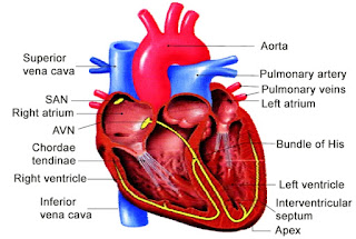
pericardium, enclosing the pericardial fluid. Our heart has four
chambers, two relatively small upper chambers called atria and two larger
lower chambers called ventricles. A thin, muscular wall called the inter-
atrial septum separates the right and the left atria, whereas a thick-walled,
the inter-ventricular septum, separates the left and the right ventricle
The atrium and the ventricle of the same side are also
separated by a thick fibrous tissue called the atrio-ventricular septum.
However, each of these septa are provided with an opening through which
the two chambers of the same side are connected. The opening between
the right atrium and the right ventricle is guarded by a valve formed of
three muscular flaps or cusps, the tricuspid valve, whereas a bicuspid
or mitral valve guards the opening between the left atrium and the left
ventricle. The openings of the right and the left ventricles into thepulmonary artery and the aorta respectively are provided with the
semilunar valves. The valves in the heart allows the flow of blood only in
one direction, i.e., from the atria to the ventricles and from the ventricles
to the pulmonary artery or aorta. These valves prevent any backward
flow.
The entire heart is made of cardiac muscles. The walls of ventricles
are much thicker than that of the atria. A specialised cardiac musculature
called the nodal tissue is also distributed in the heart
patch of this tissue is present in the right upper corner of the right atrium
called the sino-atrial node (SAN). Another mass of this tissue is seen in
the lower left corner of the right atrium close to the atrio-ventricular septum
called the atrio-ventricular node (AVN). A bundle of nodal fibres, atrio-
ventricular bundle (AV bundle) continues from the AVN which passes
through the atrio-ventricular septa to emerge on the top of the inter-
ventricular septum and immediately divides into a right and left bundle.
These branches give rise to minute fibres throughout the ventricular
musculature of the respective sides and are called purkinje fibres. The
nodal musculature has the ability to generate action potentials without
any external stimuli, i.e., it is autoexcitable. However, the number of action
potentials that could be generated in a minute vary at different parts of
the nodal system. The SAN can generate the maximum number of action
potentials, i.e., 70-75 min–1
, and is responsible for initiating and
maintaining the rhythmic contractile activity of the heart. Therefore, it is
called the pacemaker. Our heart normally beats 70-75 times in a minute
<script async src="https://pagead2.googlesyndication.com/pagead/js/adsbygoogle.js?client=ca-pub-4249528535307907"
crossorigin="anonymous"></script>


Great
ReplyDeleteKeep going ❤️
ReplyDelete